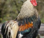
Infectious coryza is acute to subacute rapidly spreading disease of the upper respiratory tract, sinuses, and air passages of the head of several avian species that occurs worldwide. Infectious coryza is caused by the bacterium Haemophilus paragallinarum. The susceptible host for infectious coryza are chickens but occasionally it is seen in guinea fowl, turkeys, and pheasants.
Transmission occurs through direct bird-to-bird contact with clinically affected or asymptomatic carriers. This disease is transmitted through drinking water and infected suspended dust particles. It is also transmitted through indirect contact with contaminated equipment and personal. Vertical transmission is not known to occur in the infectious coryza. The incubation period ranges from 1-3 days. The route of infection is conjunctival or nasal.
Clinical signs
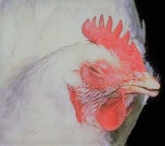
- Lacrimation and conjunctivitis (Sticking together of eyelids).
- Inflammation of eyes
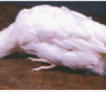
- Birds are usually lethargic having ruffled feathers and lose weight.
- Profused nasal discharge
- Sneezing, coughing, and rhinitis also occurs. Sneezing, coughing, and gurgling are indicators of tracheal and air sac involvement.
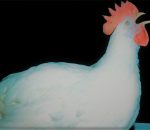
- Characteristic soft swelling of the face (facial edema). Mostly the swelling is around the eye, occasionally wattles become swollen, particularly in males.
- Egg Production is reduced by 10 to 14 percent.
- Affected broilers do not grow well and the flock appears uneven in size.
- In chronic and complicated cases of the disease, fetid odor from the flock is observed.
Post mortem lesions
- Catarrhal conjunctivitis and watery eyes
- Rhinitis
- Inflammation of the periorbital fascia. Subcutaneous edema of the face may extend to the neck and wattles.
- Congestion, petechiae in edema of the membranes of nasal passages and sinuses.
- In chronic cases, cheesy material is found in sinus cavities having a characteristic putrid odor.
Diagnosis
- Diagnosis is done on the basis of flock history and the progress of the disease.
- Also done through isolation and identification of the causative bacterium.
- Serological tests (AGPT, PCR) also assist in the process of diagnosis.
Differential diagnosis
1. Mycoplasmosis
- Mycoplasmosis or mycoplasma gallisepticum infection is slow to develop.
- A short incubation period is characteristic of infectious coryza (IP: 1-3 days).
2. Fowl cholera,
- Dead birds have liver foci in fowl cholera.
- Wattle swelling in cholera is dissimilar to swollen face in coryza.
3. Fowl pox
- Characteristic external wart-like lesions are visible in fowl pox.
- Diphtheritic lesions in the pharynx and larynx are observed in fowlpox that are absent in infectious coryza.
- Kidney lesions are absent in coryza while they are found in Infectious bronchitis (IB).
- The rate of drop in egg production in IB is more than seen in coryza.
5. Newcastle disease
- Nasal discharge is not seen in Newcastle disease (ND), while nasal discharge is present in coryza.
- Proventricular hemorrhages are seen in ND while they are absent in coryza.
6. Vitamin A deficiency
- White pimples (keratinization) on oesophageal and buccal mucosa are seen typically in vitamin A deficiency.
7. Infectious laryngotracheitis
- Bloody tracheitis is typical in ILT and this is unusual in coryza.
Prevention of coryza
- Recovered birds are resistant to re-infection. Birds recovered from the challenge of one serotype are resistant to other serotypes.
- Vaccines are used to induce immunity against the causative pathogen of coryza. Vaccines are inoculated usually at 8 to 10 weeks. Vaccines (bacterins) protect against the most severe falls in egg production although coryza may not be prevented.
- Mixing of older birds with younger birds should be discouraged as a preventive strategy. In this way, the new entries will not pick up the infection.
- Appropriate biosecurity measures should be adopted to minimize the risk of disease spreads.
- Proper flock sanitation should also be adopted. Poultry house buildings should be vacant for 30 to 60 days after cleaning and disinfecting before repopulating.
- Proper disposal of the dead birds should be managed.
- The use of preventive medication is also taken as a step for prevention.
How to manage the outbreak
- Remove all clinically affected birds and destroy or kill them. Treat the rest of the flock for at least 7 days.
- Sulphonamides are useful drugs for treatment purposes. Streptomycin intramuscular is the best choice in uncomplicated cases.
- Erythromycin reduces the spread of infection and reduces the number of shedders. Tetracyclines are also frequently used for treatment purposes.
NOTE: This disease has no public health significance reported till now.